
Signaling Pathways a Potential Target for Vascular EDS Therapies, Mouse Models Show


Investigations into two newly developed mouse models that mimic the pathology, or typical behavior of vascular Ehlers-Danlos syndrome (vEDS) have revealed that alterations in collagen production and signaling pathways underlie the connective tissue disorder.
Blocking these signaling pathways protected the mice from the rupture of the aorta artery, a severe outcome in those with vEDS.
The findings support the development of therapies to prevent vascular rupture in vEDS patients, the researchers said.
The study, “Targetable cellular signaling events mediate vascular pathology in vascular Ehlers-Danlos syndrome,” was published in the Journal of Clinical Investigation.
The vascular form of EDS, known as vEDS, is caused by mutations in the COL3A1 gene, which encodes the pro-alpha 1 chain of collagen type III, a major structural component of hollow organs such as large blood vessels, the stomach, intestines, and others.
Defective collagen III undermines the structural integrity of the supportive network outside cells — known as the extracellular matrix — which can trigger the spontaneous rupture of blood vessels or organs.
However, little is known about the underlying molecular mechanisms caused by impaired collagen III, whether signaling pathways are altered, or the effects on communication with neighboring cells.
Additionally, preclinical research into vEDS has been limited by the lack of animal models that mimic the pathology and molecular mechanisms of the disease.
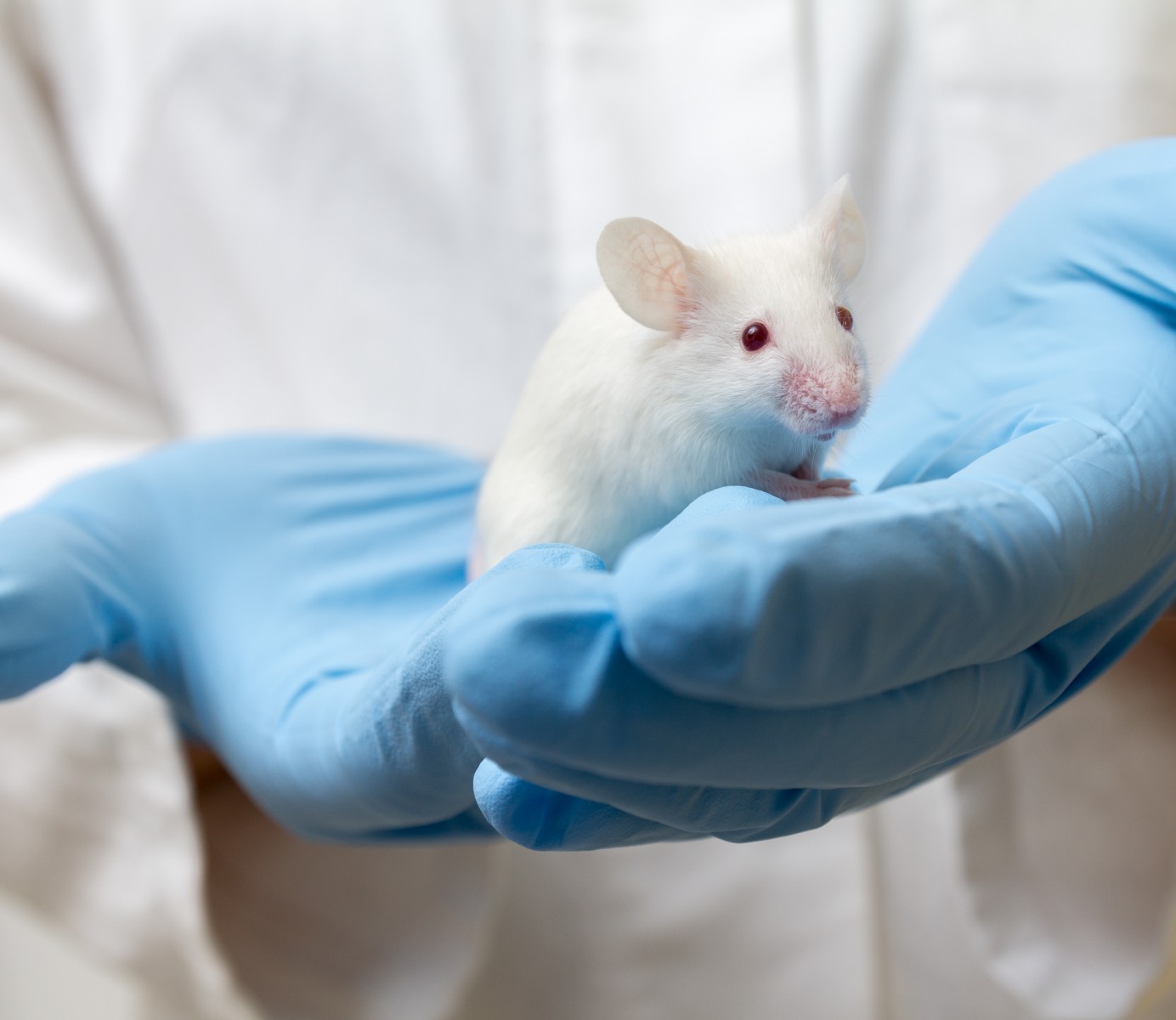
This prompted a team, led by researchers at the Johns Hopkins University School of Medicine in Baltimore, to develop two new mouse models that each carry a mutation — either Col3a1-G209S or Col3a1-G938D — found in people with vEDS, and investigate the associated molecular mechanisms leading to the disease.
Both mouse models were found to mimic vEDS characteristics in patients, with sudden death due to the rupture of the aorta — the main artery leading out of the heart.
Mice with the Col3a1-G938D mutation had more severe disease, with a median survival of 45 days, compared with 400 days for those with the Col3a1-G209S mutation.
Although the aortic wall structure was relatively preserved in both mouse models, there were minor alterations at two months of age. These alterations included elastic fiber breaks, decreased wall thickness, and less collagen content. Collagen fibrils within the aorta also showed a wide variation in diameter, and were generally smaller in size when compared with control (normal) mice.
Fibroblast cells — important for the extracellular matrix and collagen production — within the aorta showed alterations presumably due to the abnormal collagen, which also was observed in fibroblasts derived from patients.
The molecular signature for excessive signaling was found in multiple pathways, including phospholipase C (PLC), inositol 1,4,5-triphosphate (IP3), protein kinase C (PKC), and extracellular signal-regulated kinase (ERK). All these pathways are major mediators in conditions affecting the vascular system.
Interestingly, aortic rupture was prevented in mice treated with therapies that block these signaling pathways.
In mice with Col3a1-G938D mutations treated with the orally administered PKC inhibitor ruboxistaurin, 94% survived after 45 days of therapy compared with only 52% with no treatment.
Treating these mice with the approved ERK blocker cobimetinib led to the survival of 90% of mice, also after 45 days, while treatment with hydralazine resulted in 97% survival of treated mice after 45 days. An approved blood pressure medication, hydralazine works, in part, by blocking IP3.
The hydralazine protected male mice until puberty, at around 50 days, at which time there were significant rates of aortic rupture. That was consistent with the elevated risk of rupture also seen in young men with vEDS around puberty. Treatment with a medicine that blocked receptors that bind androgens (such as testosterone) prevented the ruptures.
Additionally, the use of a medicine that blocked the social bonding hormone oxytocin enhanced postpartum survival by preventing aortic rupture, a severe problem for pregnant women with vEDS.
“Taken together, our results provide evidence that targetable signaling abnormalities contribute to the pathogenesis of vEDS, highlighting unanticipated therapeutic opportunities,” the researchers said, adding that they “illustrate the promise of therapeutic strategies aimed at inhibition of the PLC/IP3/PKC/ERK axis of activation.”
“Additional work will be needed to elucidate the nature of outside-in cellular signaling that initiates with collagen III deficiency, cross-talks with androgen signaling, and culminates in PKC/ERK activation and arterial rupture in vEDS,” they added.