Family in China Has Two vEDS-causing COL3A1 Mutations
Written by |


A family in China was found to carry two mutations causing vascular Ehlers-Danlos syndrome (vEDS) over multiple generations, a study reports.
The mutations interacted such that their effect was beyond that of each mutation alone. “This is the first report of such a combination ever described to cause vEDS in the literature,” the scientists wrote.
The study, “Two closely spaced missense COL3A1 variants in cis cause vascular Ehlers-Danlos syndrome in one large Chinese family,” was published in the Journal of Cellular and Molecular Biology.
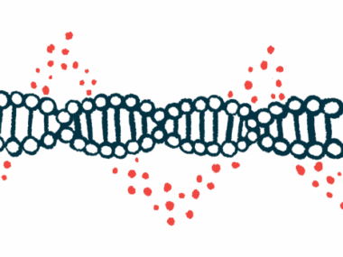
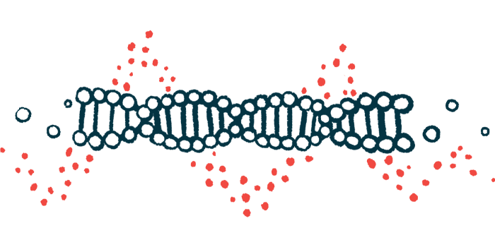
The vascular type of EDS is characterized by fragile arteries and internal organs, as well as thin, translucent skin that bruises easily. It is typically caused by mutations in the COL3A1 gene. This gene provides instructions for a portion of type 3 collagen, a protein present in the connective tissue that holds tissues together.
In this study, a team of researchers described a family that was found to carry two disease-causing COL3A1 mutations over multiple generations.
The first member of the family to bring the case to the doctors’ attention (the proband) was a 36-year-old man who went to the hospital because of pain in the upper abdomen. A physical exam revealed velvety and soft skin with veins that could be seen through it in the hands and feet. He also showed an aged appearance in the extremities (known as acrogeria) and bruises caused by blood leaking from broken blood vessels into the skin (ecchymosis).
A CT angiography scan to provide pictures of the blood vessels revealed aneurysms of certain arteries. An aneurysm is a bulge in an artery resulting from a weakness in its wall.
When asked about his family’s history, the patient said that his grandmother, father, and two uncles had died suddenly at age 25–48 of unexplained abdomen pain, headache, or nosebleed.
Based on this information, all living blood relatives were called for a clinical examination.
A 31-year-old male cousin had unusually stretchy skin, ecchymosis, scarring, and hypermobile joints (moving beyond the normal range of movement). A 39-year-old sister had a tear in the inner layer of the aorta, a large artery that carries oxygen-rich blood away from the heart to the rest of the body. She also had an aneurysm in the abdominal portion of the aorta. A 41-year-old female cousin had a ruptured artery in the brain.
The other blood relatives appeared healthy.
To search for a common cause of the symptoms, the doctors performed genetic testing on the proband for a panel of eight genes associated with EDS. They found two closely spaced mutations in COL3A1: one was p.Leu734Phe (c.2199_2200TC>AT), which had never been reported before, and the other was p.Gly741Ser (c.2221G>A), a previously known mutation.
Next, the researchers checked whether the other family members also carried the mutations in COL3A1. Of the 19 family members tested, six carried both mutations.
“To the best of our knowledge, such a large and well-informed vEDS family has never been reported in the literature,” the researchers wrote.
The mutations were found not only in clinically affected family members but also in two healthy relatives (a baby less than 1 year old and a 16-year-old boy).
“In this regard, it is worth mentioning that in a European cohort study, only 17% of vEDS patients had experienced an initial complication by the age of 20,” the scientists wrote, adding that the two children should receive genetic counseling to help the family understand the risks of carrying the mutations.
To know if the mutations affected the COL3A1 protein that was produced, the researchers went to the lab and introduced the gene into skin fibroblasts, which are cells of the connective tissue that make collagen proteins. The team used either the healthy (wild-type) copy of the gene or one that contains either of them or a combination of the two mutations. Then, they watched for changes in the levels of COL3A1.
Compared with the wild-type gene, each mutation independently led to a lower COL3A1 level and, when combined, to an even more reduced COL3A1 amount.
Based on these findings, each of the mutations can be classified as causing disease, yet their effect was greater when occurring together, the investigators said. “We are not aware of any similar findings in the literature,” they wrote.