
Overlooked CT scan clue shows cerebral air embolism with vEDS
The condition is caused by air entering blood vessels in the brain
Written by |


A young man with vascular Ehlers-Danlos syndrome (EDS) developed a cerebral air embolism caused by air entering blood vessels in his brain that was difficult to diagnose and illustrated the importance of carefully reviewing CT scans.
“Prompt identification of cerebral air embolism allows supportive measures to be started to prevent potentially fatal sequelae,” researchers in the U.K. wrote in “Cerebral air embolism in vascular Ehlers-Danlos syndrome: a retrospective diagnosis,” which appeared in Practical Neurology.
To treat a cerebral air embolism, the patient should be placed in the Trendelenburg position — lying on the back with the head lower than the feet — to keep air bubbles away from the heart’s main outflow. At the same time, the patient should receive 100% oxygen at a high flow.
EDS is a group of genetic diseases that affect connective tissues. As a result, patients may have overly flexible joints and thin, fragile skin that’s unusually stretchy. Along with these symptoms, people with vascular EDS also have fragile internal organs and blood vessels that are prone to rupturing, causing serious complications.

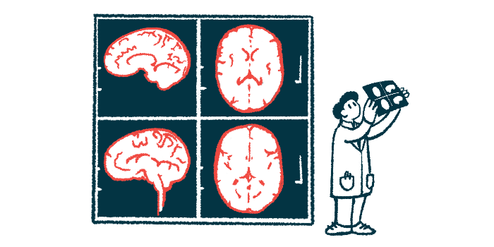
CT scan review helps correct misdiagnosis
Here, researchers described the case of a 24-year-old man with reduced consciousness who was agitated, didn’t respond to instructions, and wasn’t moving the left side of his body when he was examined at a hospital. His breathing was quick at 24 breaths a minute and he needed oxygen supplementation.
For three days, he’d had a mild headache on the right side and coughed up small amounts of blood. His mother noticed a change in his behavior, including agitation, silence, uncontrolled limb movements, and loss of bladder control. He had a history of autism and a genetically confirmed diagnosis of vascular EDS. His EDS symptoms included flexible joints, bruising, repeated tears in the main blood vessels in the neck, migraine, and collapsed lungs caused by leaked air, called pneumothorax.
CT scans showed signs of infection in his right lung and a small bleed in the right side of his brain. He was started on the antiviral aciclovir and broad-spectrum antibiotics in case he had an infection in either one.
A week after he arrived at the hospital, an MRI showed several areas of damage in the outer layer of his brain, mostly on the right side, but also affecting the front and back. There was also damage in the middle part of his brain. Small areas of bleeding were seen near where the earlier bleed had occurred and a separate scan showed a blockage caused by a tear in the right vertebral artery, leading the doctors to consider a diagnosis of posterior reversible encephalopathy syndrome.
After the man was discharged, a review of his CT scan revealed a pocket of air inside the bleed on the right side of his brain. A CT scan obtained 24 hours later showed the air had disappeared. The doctors later diagnosed him with a cerebral air embolism. Signs of bleeding in the lungs as well as a bronchovenous fistula — an abnormal connection between an airway and a vein that allows air to enter the bloodstream — were also seen after the original CT scan of the chest was rechecked.
The man was being followed by lung specialists, who may consider blocking the bleeding blood vessel, at the time the study concluded.
“This patient was incorrectly diagnosed with posterior reversible encephalopathy syndrome before subsequent closer inspection of the CT scan at presentation disclosed the true diagnosis,” the researchers wrote. “The likely cause in this case was a transient bronchovenous fistula associated with vascular Ehlers-Danlos syndrome.”