
Case of Vascular EDS, Recurrent Pneumothorax, Pulmonary Complications Described in Report
Written by |


A patient with vascular Ehlers-Danlos syndrome (vEDS), recurrent pneumothorax, and other atypical pulmonary complications was described in a case report from South Korea.
The case study, “Vascular Ehlers-Danlos syndrome with cryptorchidism, recurrent pneumothorax, and pulmonary capillary hemangiomatosis-like foci, A case report,” was published in the journal Medicine.
Vascular EDS is one type of Ehlers-Danlos syndrome and is characterized by thin, transparent skin, moderate hyperflexibility of small joints, and fragility of vital organs and blood vessels.
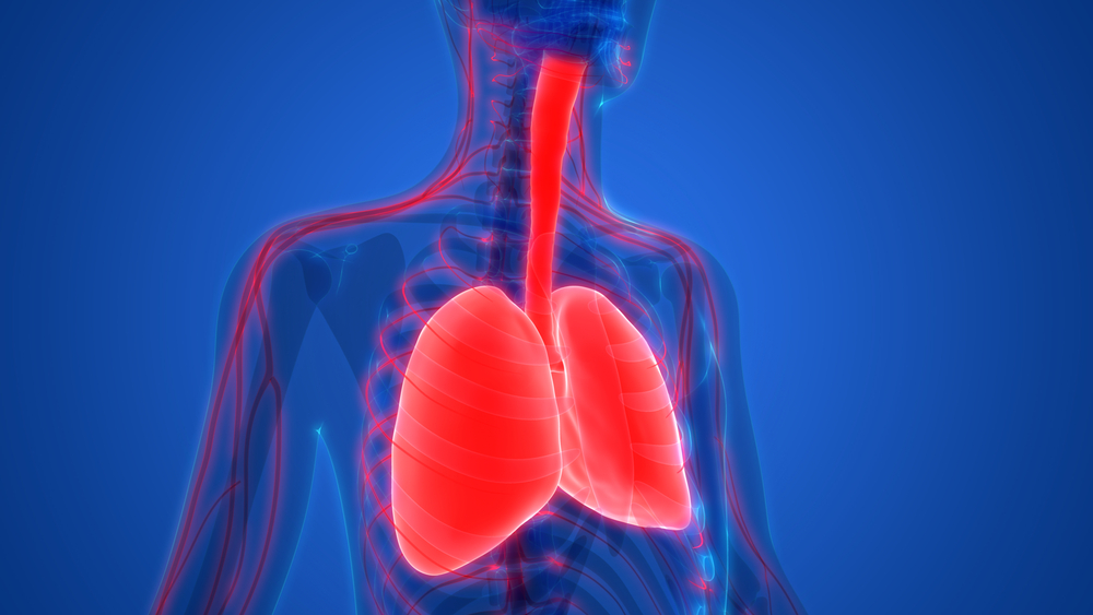
Pulmonary complications, including pneumothorax — a collapsed lung due to air leaking into the space between the lung and chest wall — have been reported in vEDS patients, especially in young adults.
However, no vEDS studies have described the occurrence of pulmonary capillary hemangiomatosis (PCH), a rare cause of pulmonary hypertension characterized by the proliferation of pulmonary capillaries.
Now, researchers described the case of an 18-year-old teen male with chest pain and dyspnea, or shortness of breath. The patient had two cases of spontaneous pneumothorax, the first on the right side and the second on the left, treated with tube thoracostomy, which involves inserting a tube into the pleural cavity to drain fluid.
He had undergone surgeries to correct a deviated septum in his nose and a cryptorchidism, which happens when one of the testes fails to descend into the scrotum, on the right side. The young man had no history of tobacco smoking or any significant familial disease.
A chest auscultation revealed reduced lung sound on the right side. Levels of serum creatinine (an indicator of liver function), white blood cells, platelets, and hemoglobin were normal.
On a chest X-ray, clinicians found a right-sided pneumothorax. He then underwent a video-assisted bullectomy — surgery to remove a dilated air space in the lung — for his recurrent pneumothorax.
However, air leakage persisted, which led the physicians to perform a pleurodesis, a surgical procedure that seals the space between chest walls and lungs to eliminate fluid buildup.
The patient’s air leakage remained, even after additional pleurodesis.
A chest computed tomography (CT) scan revealed hyperinflated lungs, abnormal, with low attenuation (density) in the lung parenchyma — the portion of the lung involved in gas exchange — and new cystic lesions.
Compared to CT scans done three and seven months earlier, areas of increased attenuation were more pronounced, and cystic lesions were newly developed.
Biochemical tests for autoantibodies were negative. However, the levels of both red and white blood cells suggested intrapulmonary hemorrhage.
During a second video-assisted lung biopsy, the team observed hemorrhage-like changes and several bruise-like lesions in the lung parenchyma.
Capillary proliferation with pleural fibrosis (scarring) and macrophages — a type of immune cell — within the lung’s tiny air sacs (alveoli) were also found. This was consistent with a diagnosis of PCH. However, no evidence of pulmonary hypertension or cardiac abnormality was detected.
“Thus, we concluded that the lung lesions were not PCH, but PCH-like foci,” the team wrote.
Aiming to identify the cause of the atypical manifestations, the clinicians conducted additional tests. The young man was thin, had hypermobile joints especially in his fingers and wrists, as well as hyperextensible and transparent skin, which, combined with the recurrent pneumothorax, suggested EDS.
Upon finding an extremely rare mutation in the COL3A1 gene, a final diagnosis of vEDS with PCH-like foci and cryptorchidism was made.
The team could not determine whether the mutation was sporadic or inherited, because the patient’s parents refused to undergo genetic tests.
Of note, no reports of cryptorchidism in vEDS had been published, the researchers said.
“To the best of our knowledge, both cryptorchidism and PCH-like foci have never been reported yet as complications of vEDS, suggesting our case might be a new variant of this condition,” they wrote.
Pulmonary function results before the patient was discharged were compatible with emphysema-related change and bronchial asthma, but no asthma symptoms were detected. Ten months after discharge, the patient remained symptom-free, with no relapse of pneumothorax, new lesions, or pulmonary hypertension.
“This case emphasizes the importance of comprehensive physical examination and history-taking, and the clinical suspicion of a possible connective tissue disorder when we encounter cases with atypical presentation and/or unique chest radiologic findings especially in young patients,” the researchers wrote.
“It is important for his family to be informed about the future risk of vascular or surgical complications due to fragile tissues, the necessity of close follow-up for the potentially fatal complications of the condition, and be strongly encouraged to receive genetic counseling,” they added.


Leave a comment
Fill in the required fields to post. Your email address will not be published.