
Case Study: Check for vEDS in Patients With Pneumothorax
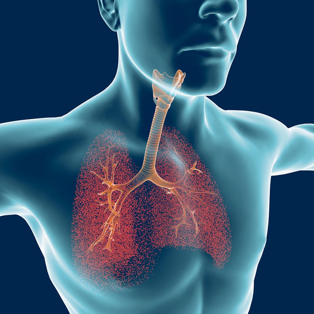
Naeblys/Shutterstock lungs


Doctors should consider the possibility of vascular Ehlers–Danlos syndrome (EDS) when seeing a patient with recurrent pneumothorax — the presence of air in the space between the lungs and the chest wall — that’s resistant to conventional therapy, according to a case report.
The report described a 19-year-old man who was diagnosed with vascular EDS, or vEDS, only after repeated hospitalizations.
“This case highlights the importance of detailed history-taking, physical examination, and clinical suspicion of vascular EDS when encountering recurrent pneumothorax cases refractory [resistant] to conventional therapy,” the researchers wrote.
The report, “Ehlers–Danlos syndrome presenting as cystic lung disease with recurrent pneumothorax: a case report,” was published in the journal Respirology Case Reports.

Vascular EDS is characterized by fragile arteries, muscles, and internal organs, and thin, translucent skin that bruises easily. Pneumothorax is another common finding in people with vEDS.
Here, researchers from the University of Ulsan College of Medicine, in Seoul, Korea, described the case of a man with lung cysts, or sacs of tissue filled with air or fluid. The patient had experienced several episodes of pneumothorax, either in the right or left lung, which were resistant to widely used surgical procedures such as closed thoracotomy — to gain access to the lungs — and wedge resection, done to remove a triangle-shaped slice of tissue.
Because of repeated pneumothorax, the young man was referred to another hospital for further evaluation and diagnosis.
On admission, a chest CT scan showed a pneumothorax in the right lung and multiple ground-glass opacities, a radiologic feature seen in various clinical conditions. The patient also was found to have nodules — small growths — in both lungs.
A detailed history revealed that the patient coughed up blood whenever pneumothorax developed. He also had a history of easy bruising and a family history of sudden death, with his father having died of a brain bleed. He had no history of smoking.
A physical exam revealed deep-set eyes, and joints that were hypermobile, meaning they stretch beyond normal limits. His skin was thin with visible blood vessels and he bruised easily.
Based on these findings, doctors suspected the patient had vEDS as per the current 2017 classification system.
To confirm the suspicion, the team performed genetic testing to screen for mutations in COL3A1, a gene encoding type 3 collagen that is linked to vEDS. Type 3 collagen is a protein that supports and strengthens the body’s tissues and organs together.
A mutation, c.1662+1 G>A, was found in one of the two gene copies. This mutation already had been reported to cause vascular EDS.
After the diagnosis, the patient was further evaluated for arterial complications resulting from the disease. However, all arteries were normal. He was being monitored on an outpatient basis as of the study’s completion.
While “recommendations for the management of pneumothorax in vascular EDS have not been made … a conservative approach is recommended as much as possible,” the team wrote. Unless needed, surgical procedures should be avoided due to the risk of bleeding, they noted.