Vascular EDS found in 2 adults treated for serious eye problems
Complications were life-threatening for man and woman in Romania
Written by |


Two cases of vascular Ehlers-Danlos syndrome (vEDS) were diagnosed during the treatment of severe eye-related problems and later found to be vEDS-associated complications, according to a recent report from Romania.
While these complications were considered life-threatening for the young adults, prompt identification of their underlying cause enabled both patients to receive appropriate treatment.
“Each patient may have different clinical aspects that require different approaches, so being aware of all sides of this pathology and keeping an open mind as clinicians could make a difference in these cases, regardless of the gravity of their clinical state,” the scientists wrote.
The case report, “Uncommon association between vascular Ehlers–Danlos syndrome and ocular complications,” was published in the journal Frontiers in Medicine.

vEDS marked by fragile blood vessels prone to tears and ruptures
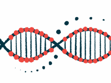
Ehlers-Danlos syndrome (EDS) refers to a group of genetic diseases characterized by weakness in the body’s connective tissues. vEDS is a severe type of EDS typically caused by mutations in the COL3A1 gene.
In addition to common EDS symptoms like overly mobile joints and fragile skin, vEDS is marked by weak and fragile blood vessels that are prone to tearing or rupturing, leading to potentially serious internal bleeding, in addition to ruptures in organs like the lungs or colon.
When blood vessels in the eyes are impacted, vEDS patients might experience visual or other ocular symptoms, such as blurred vision, dry eyes, or headaches.
Scientists report the cases of two adults with vEDS associated with serious eye problems, who were referred to their clinic in Bucharest.
A 28-year-old woman came to a hospital’s emergency room with complaints of double vision, eye bulging, headache, ringing in the ears, and vertigo, or a feeling that the room is spinning. The symptoms had lasted a few days and were accompanied by nausea and vomiting.
At the hospital, she began experiencing intense back pain, and an exam found low blood pressure, a fast heart rate, and signs of hemorrhagic shock, a severe response to bleeding that leads to inadequate oxygen delivery to the body’s cells.
An aneurysm — an abnormal bulge in a blood vessel wall — was identified in an artery of the kidney, as well as signs of internal bleeding and compromised blood flow to the left kidney. The woman underwent emergency surgery, but continued with acute kidney failure that required intervention.
Once stable, she was transferred to the Bucharest hospital on suspicion of a right-sided carotid cavernous fistula, or an abnormal connection between the arteries that run up the neck and the collection of veins located behind the eyes.
She had low visual acuity and high pressure in that eye, in addition to a number of other ocular abnormalities. It was also noted that the woman had a narrow nose, translucent skin, and thin lips, all of which can be signs of EDS.
The presence of the fistula was confirmed, as was stenosis, or narrowing, and an abnormal appearance of some arteries. She underwent a procedure that corrected the fistula.
Given that several diagnostic criteria for vEDS were met, she was given genetic testing that confirmed the presence of a mutation in COL3A1.
The other patient, a 28-year-old man, came to the emergency room with a sudden loss of vision beginning two weeks earlier.
As with the previous case, the man exhibited signs of vEDS, including translucent skin, a narrow nose, and thin lips. An eye exam revealed normal visual acuity in the right eye, but no central vision in the left eye.
He ultimately was found to have an occlusion, or blockage, in the central retinal artery — a blood vessel that supplies blood to the part of the eye that’s critical for vision.
Additional tests found signs of vEDS in the arteries, including tears, stenosis, and other anatomical changes.
He underwent surgeries to restore more normal blood flow. Again, genetic testing confirmed a mutation in COL3A1.
The man was treated with anticoagulants to prevent blood clots and advised to avoid significant physical exertion that can increase the risk of artery tears.
“Diagnosing vEDS is not an easy task,” the scientists wrote. “It is important to recognize the signs of such [a] disease, since the right diagnosis, treatment, and prevention of further lesions, may make the difference between life and death.”